Stories
- Article
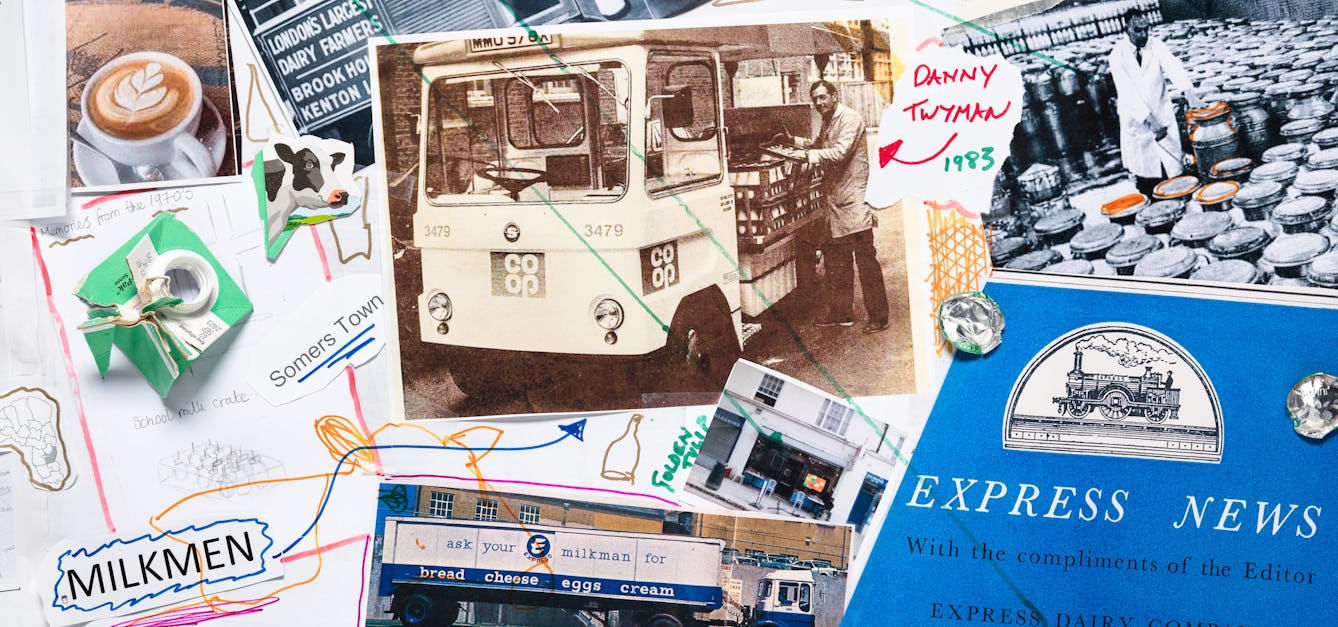
Milk trails round Euston
Where cows once grazed near Wellcome Collection in London, baristas now froth their milk. Esther Leslie uncovers Euston’s dairy-based urban history.

- Article
Coasting to catastrophe
In climate change, everything – and everyone – is connected. The watery process that will gradually cut off the Isle of Thanet from the British mainland has begun, and everyone in the UK needs to pay attention.

- Article
The amateur silversmith
It started as hobby and soon became a passion. Geraldine Holden tells us where the art and science of silver unite.
- Book extract
Eating their own kind
In his grisly history of cannibalism, zoologist Bill Schutt asks what drives an animal to feast on its own flesh and blood.


Catalogue
- Archives and manuscripts
`Illustrations and graphs of my calculations for capillary surface area (not published)'
Date: Mid 20th centuryReference: PP/DBD/B/30Part of: Daly, Professor Ivan de Burgh- Books
Patna City municipality, surface drainage construction : area blocks 36 and 37 : report / [F. C. Temple].
Temple, F. C. (Frederick Charles)Date: [1919]- Books
Possible relations of the weight of the lungs and other organs to body-weight and surface area (in dogs) / by G.N. Stewart.
Stewart, G. N. (George Neil), 1860-1930.Date: [1921?]
- Digital Images
- Online
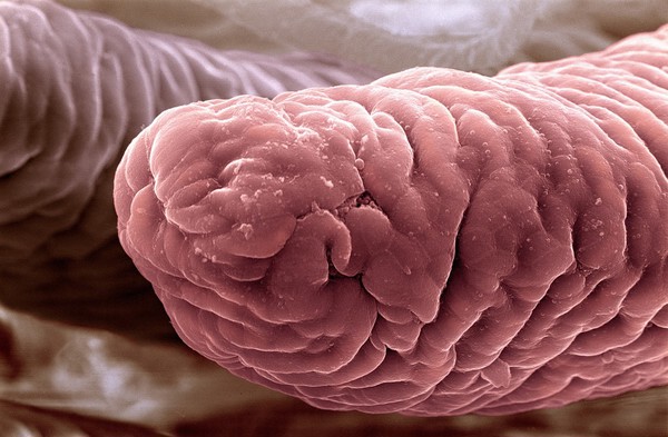
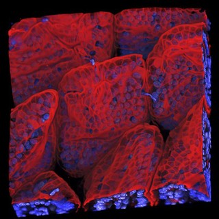
Inner surface of ileum
Prof Giorgio Gabella
- Digital Images
- Online
Leucoma on the tongue with fluffy surface
Godart, Thomas